


Radiology

Volumetric Computed Tomography (VCT) scans have revolutionized medical imaging for humans, providing detailed 3D images of the body with minimal discomfort. However, when it comes to our furry friends, VCT scans aren’t typically used. Here’s why: 1) Anatomical Differences The primary reason VCT scans aren’t used on dogs is due to the significant anatomical differences between humans and dogs. VCT scanners are calibrated and designed to capture human body structures accurately. Dogs, on the other hand, have different bone densities, organ placements, and overall body structures that might not be accurately captured by a VCT scanner designed for humans. This can lead to less precise images and potentially inaccurate diagnoses. 2) Size and Positioning VCT scanners are built to accommodate the size and positioning of human patients. Humans generally have a standard size and shape that these machines are designed to handle. Dogs, however, come in a wide variety of sizes and shapes, from tiny Chihuahuas to large Great Danes. Positioning a dog correctly in a machine designed for humans can be challenging and may not yield the best results. 3) Motion and Sedation One of the key requirements for a successful VCT scan is that the patient remains still during the procedure. While humans can follow instructions to stay still, dogs often need to be sedated to achieve the same level of stillness. Sedation in animals can be more complex and carries its own risks, especially in a machine not designed for their use. This adds an additional layer of complexity to using VCT scans on dogs. 4) Specialized Veterinary Equipment Veterinary medicine has its own set of specialized imaging equipment, such as veterinary-specific CT and MRI machines. These machines are tailored to the needs of animals and are designed to capture the unique anatomical details of dogs and other animals. They provide the necessary adjustments and calibrations to ensure accurate and safe imaging for veterinary patients. 5) Radiation Safety The radiation dose and safety protocols for VCT scans are established based on human studies. Using these machines on dogs without proper adjustments could pose unnecessary risks. Veterinary-specific imaging equipment is designed with the appropriate safety measures for animals, ensuring that they receive the correct amount of radiation for accurate imaging without undue risk. BUT WHAT CAN WE USE TO SCAN OUR FURRY FRIENDS?! There are several imaging techniques specifically designed for veterinary use that can effectively scan dogs. Here are some of the most common ones: 1. X-rays (Radiography) X-rays are widely used in veterinary practices to create images of bones, organs, and other internal structures. They are particularly useful for detecting fractures, tumors, and foreign objects . 2. Ultrasound (Ultrasonography) Ultrasound uses sound waves to create images of the inside of the body. It’s commonly used to examine soft tissues, such as organs and blood vessels, and is particularly useful for diagnosing conditions like heart disease and abdominal issues . 3. CT Scans (Computed Tomography) CT scans provide detailed cross-sectional images of the body and are useful for diagnosing complex conditions, such as tumors, internal injuries, and bone disorders. Veterinary-specific CT scanners are designed to accommodate the anatomical differences of animals . 4. MRI (Magnetic Resonance Imaging) MRI uses magnetic fields and radio waves to produce detailed images of soft tissues, such as the brain, spinal cord, and muscles. It’s particularly useful for diagnosing neurological conditions and soft tissue injuries . Like CT scans, MRIs require the dog to be sedated or anesthetized to ensure they remain still during the procedure. 5. Nuclear Medicine This technique involves the use of small amounts of radioactive material to diagnose and treat diseases. It can provide information about the function of organs and tissues, which is useful for diagnosing conditions like cancer and thyroid disorders . Conclusion While VCT technology is advanced and highly effective for human diagnostics, veterinary medicine relies on equipment specifically designed for animals to ensure accurate and safe imaging. The anatomical differences, size and positioning challenges, need for sedation, and radiation safety concerns all contribute to why VCT scans aren’t typically used on dogs. Instead, veterinarians use specialized imaging tools that are better suited to capture the unique details of our beloved pets, ensuring they receive the best possible care.
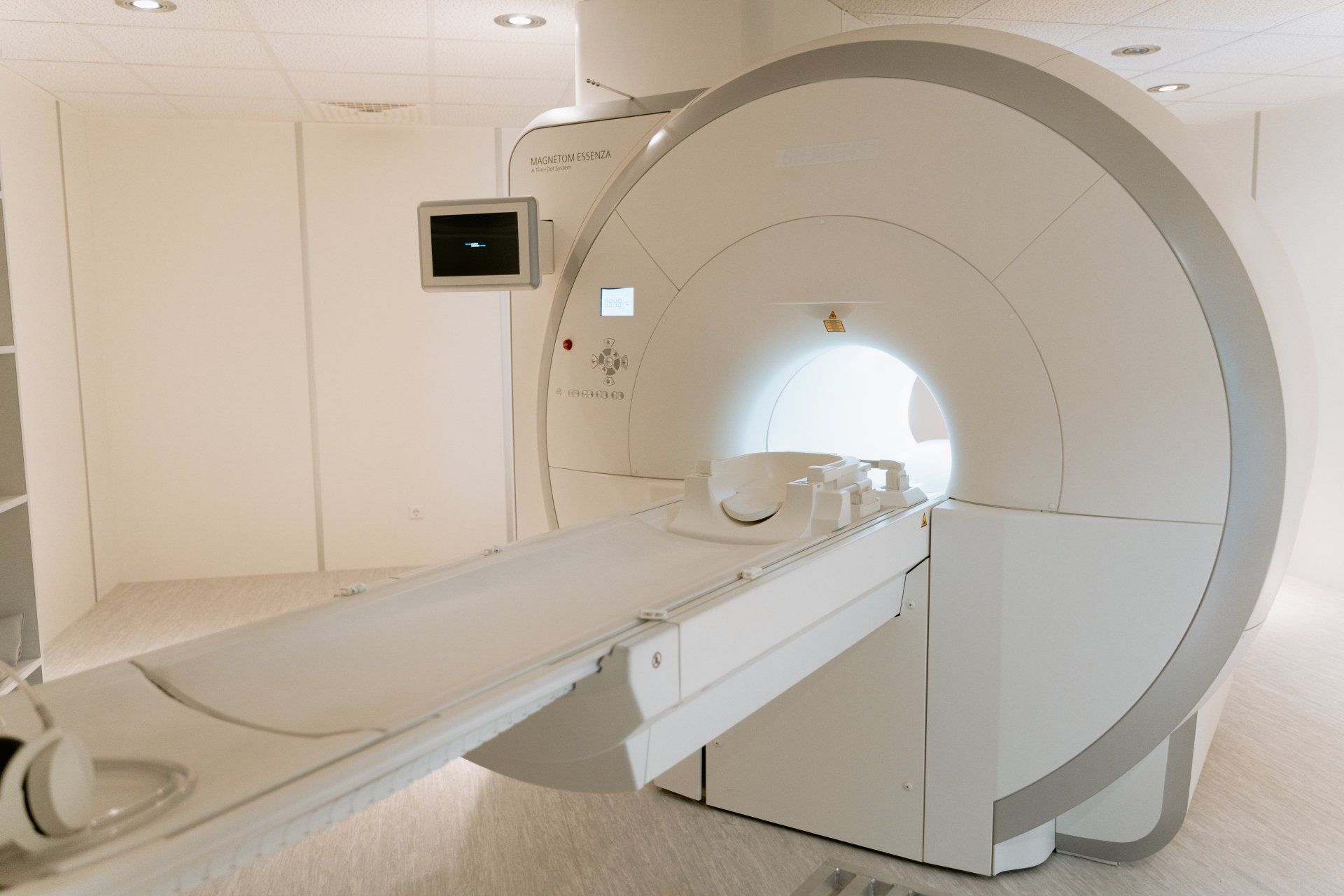
After a lot of experimentation, scientists have developed a new method of imaging that captures 3D images of the human body using Volumetric Computed Tomography (VCT). Not only that, but it has kept improving and evolving… VCT scanners are now more powerful, quick, and stylish than ever. For anyone who has ever had to remain motionless for longer than five minutes (which is pretty much everyone), the fact that they can scan your body in a matter of seconds is fantastic news. VCT has come a long way, transforming from bulky and slow to sleek and fast. So, how does Volumetric Computed Tomography (VCT), a cutting-edge imaging technique, provide high-resolution, three-dimensional images of the human body while limiting radiation exposure and keeping patients comfortable and safe during the scan? This is the most amazing question to address first. Here’s how it works: As you gently pass through a large doughnut-shaped machine while lying down on a table, don’t be alarmed; it’s not as scary as it sounds. The machine continuously takes x-rays of your body from various angles as you pass through it. These images are then transferred to a computer, which assembles them into a 3D model of your body. The good news is that VCT scans are rapid and comfortable! There are no uncomfortable elements, such as radiation or sticks, to worry about. Plus, the images are incredibly detailed. Certainly! There are numerous applications for Volumetric Computed Tomography (VCT) scans in various industries, including: Medical diagnosis : VCT can detect and diagnose a variety of medical conditions, including cancer, heart disease, and neurological disorders. Treatment planning : VCT allows clinicians to plan and prepare for surgical procedures, radiation therapy, and other types of medical treatments by obtaining precise 3D images of the body. Monitoring illness progression : VCT can be used to track changes in the body over time, helping medical professionals monitor the progression of certain diseases and assess the effectiveness of treatments. Research : VCT can be used to develop and test new medical technologies and therapies, as well as study the anatomy and function of the human body. So, you might ask yourself how precise and reliable the 3D images generated by Volumetric Computed Tomography (VCT) scans can be… VCT scans produce accurate, dependable, and highly detailed 3D images of the body. Bones, organs, blood vessels, and tissues are all incredibly clear for doctors to observe. It’s like having a clear window into the inner workings of the body without having to undergo any invasive procedures. Therefore, you can be confident that if you require a VCT scan, it will provide your doctor with incredibly accurate and useful information to help with your medical diagnosis and treatment. What about the risks associated with this machine?? VCT (Volumetric Computed Tomography) scans are typically regarded as safe, non-invasive, and have few adverse effects. However, there are a few potential risks and considerations to keep in mind, such as: Radiation exposure : Ionizing radiation, which is present during VCT scans, may occasionally raise cancer risk. A single VCT scan normally exposes the patient to a small amount of radiation, which is unlikely to cause harm. Allergies : Some individuals may develop allergies to the contrast dye used in VCT scans. Symptoms can include hives, breathing problems, and itching. Although rare, this can sometimes be treated with medication. Claustrophobia : Some people may feel anxious or claustrophobic during the VCT scan, as it involves lying still in a narrow tube for several minutes. However, this can often be managed with relaxation techniques or medication to help you feel more comfortable. Pregnancy : VCT scans are generally not recommended for pregnant women, as they involve exposure to ionizing radiation that can potentially harm the developing fetus. If a VCT scan is necessary during pregnancy, your doctor will take precautions to minimize radiation exposure to the fetus. Overall, while VCT scans are generally safe and well-tolerated, it’s important to discuss any potential risks and concerns with your doctor before undergoing the procedure. Fun fact about VCT scans : No matter how sophisticated VCT technology may be, it still can’t be used for dogs and is only applicable to human patients. So, for the time being, we’ll just have to settle for standard dog X-rays. Check this article for more details on why it can't be used for dogs 😆 .
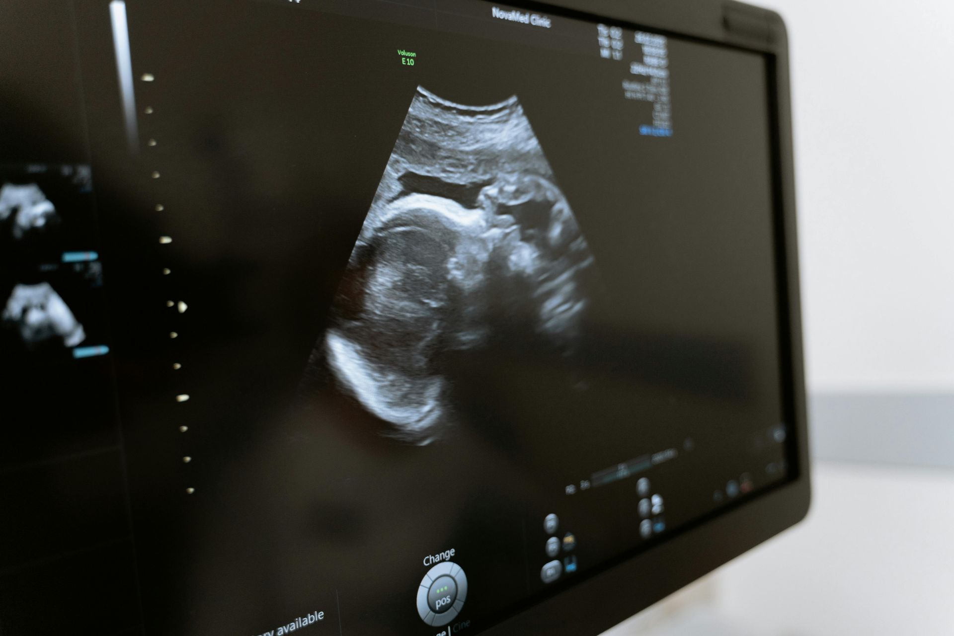
The story of ultrasound exploration is one of invention and perseverance, driven by an attempt to discover the secrets of the human body. Researchers have pushed the boundaries of science to explore the potential of sound waves for medical imaging, from the creation of sonar technology during World War I and II through the pioneering work of Karl Dussik in Austria. This road of discovery has resulted in a revolution in medical practice, with ultrasound now being used to diagnose and treat a wide range of diseases. Join us on a fascinating tour through the history of ultrasound to see how this extraordinary technology has changed the appearance of modern medicine. To begin, understanding how ultrasound works will help us appreciate the incredible power and promise of this remarkable technology. So, to begin, consider the following question: What is the procedure of an ultrasound?
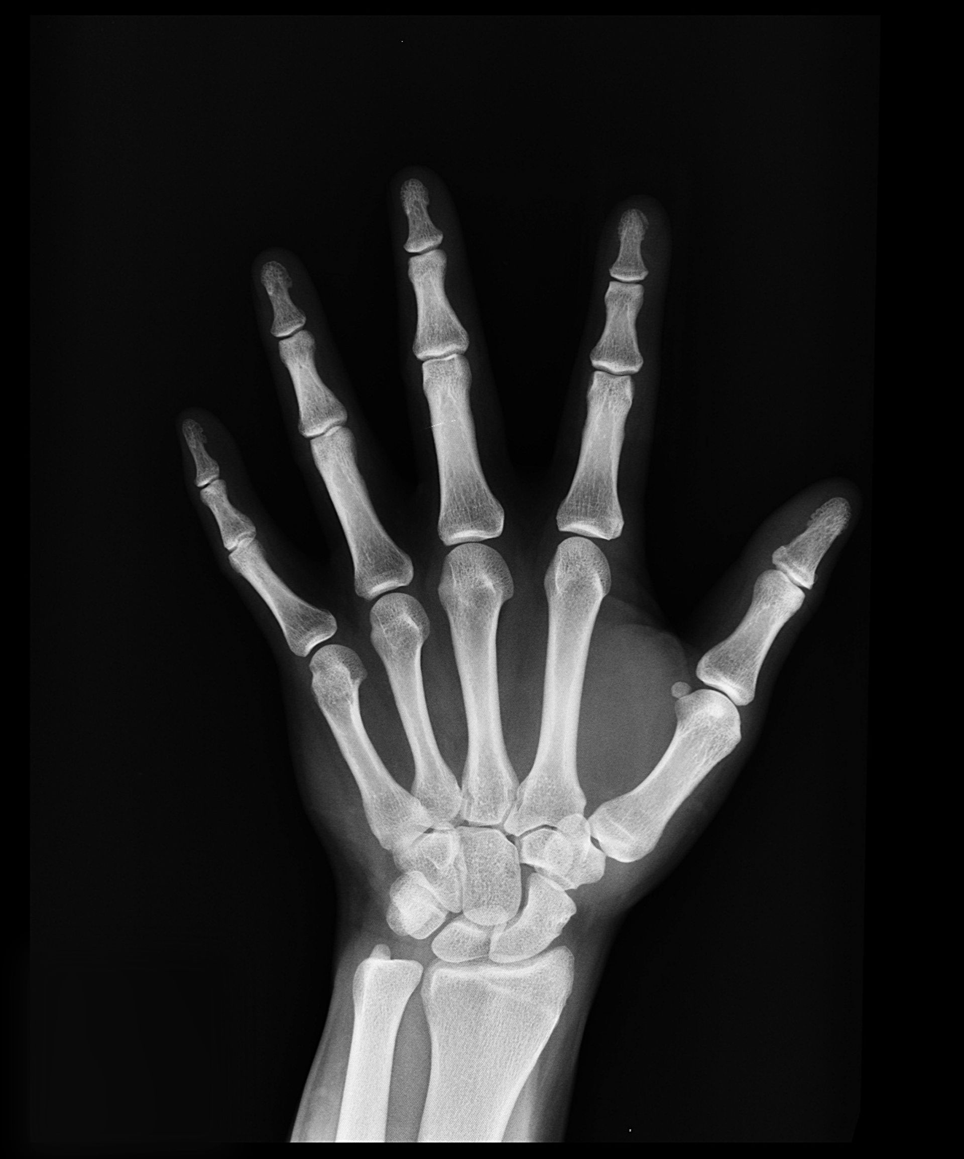
These strong rays have been used by doctors and scientists for almost a century to gaze into the mysteries of the human body and beyond. But did you know that X-rays were found entirely by chance ? In 1895, Wilhelm Conrad Roentgen, a German physicist, was experimenting with cathode rays (aka electric currents) when he noticed an unusual glow coming from a nearby screen. He had no idea that this unintentional discovery would forever revolutionize the world of medicine! Radiation such as X-rays can travel through the body. They are invisible to the naked eye and cannot be felt. The energy from X-rays is absorbed in varying rates by different regions of the body as they move through the body. After the X-rays have passed through, a detector on the other side of the body collects them and converts them into an image. Dense portions of your body, such as bone, that X-rays find difficult to get through, appear as distinct white patches in the image. Softer tissues that X-rays can easily pass through, such as your heart and lungs, seem darker. So, what’s the true purpose of X-rays? They are commonly used for a variety of evaluations, of which we shall highlight a few: Dental X-rays are used in dentistry to obtain images of the teeth and surrounding tissues, which aid in the diagnosis of oral health concerns and treatment planning. Cancer Treatment: a type of radiation therapy that uses high doses of radiation to eliminate cancer cells in specific parts of the body. Veterinary Care: utilized in veterinary medicine to diagnose and treat animals, assisting veterinarians in the detection of fractures, cancers, and other health problems. Industrial inspection is used to inspect machinery and infrastructure in the industrial sector, assisting in the detection of flaws and the prevention of accidents. Forensic Science: a branch of forensic science that examines the evidence and reconstructs crime scenes to assist in identifying victims of crimes and accidents. Bone Density Testing: used to assess bone density and aid in the diagnosis of osteoporosis and other bone-related diseases. The chart below provides additional information about the medical applications of X-rays: